

At a Glance
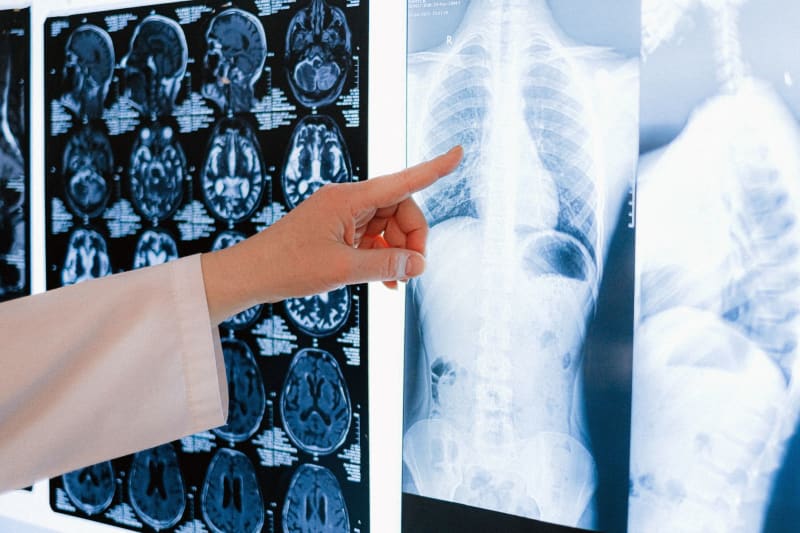
- Researchers at the National Eye Institute (NEI) have developed a novel MRI technique, significantly enhancing the resolution and clarity of brain and eye imaging, promising a new era in diagnostic capabilities.
- This advanced MRI system achieves an unprecedented level of detail, allowing scientists to visualize structures within the eye and brain that were previously undetectable with conventional imaging methods, opening new avenues for research and clinical application.
- The breakthrough involves a specialized coil and optimized pulse sequences, specifically engineered to overcome the inherent challenges of imaging delicate ocular tissues and intricate neural pathways.
- Early applications of this technology are focused on understanding the progression of age-related macular degeneration (AMD) and other complex neurodegenerative diseases, offering a clearer picture of their underlying mechanisms.
- This innovation holds immense potential for earlier and more precise diagnosis of various conditions, including glaucoma, diabetic retinopathy, and even subtle changes indicative of Alzheimer's disease or multiple sclerosis.
- The enhanced imaging capabilities are expected to accelerate the development and evaluation of new therapeutic interventions, providing objective metrics for treatment efficacy and patient response.
The Record
For decades, Magnetic Resonance Imaging (MRI) has been a cornerstone of medical diagnostics, offering non-invasive insights into the human body's soft tissues. However, imaging the eye and certain intricate brain regions with sufficient clarity has remained a persistent challenge. The delicate, microscopic structures of the retina, optic nerve, and specific brain nuclei often blur into indistinctness with standard MRI protocols, limiting their diagnostic utility for early-stage diseases or subtle pathological changes. This technological hurdle has meant that many conditions affecting vision and neurological function could only be diagnosed at later stages, often after significant, irreversible damage had occurred, underscoring the critical need for higher-resolution imaging.
The National Eye Institute (NEI), a division of the National Institutes of Health, has been at the forefront of ophthalmic research for over 50 years, consistently pushing the boundaries of understanding and treating eye diseases. Their mission extends beyond basic science to the development of translational technologies that directly impact patient care. This latest MRI breakthrough is a testament to their sustained commitment to innovation, building upon years of foundational research in imaging science and neuro-ophthalmology. The institute's collaborative environment, bringing together physicists, engineers, and clinicians, has been instrumental in transforming theoretical concepts into practical, high-impact diagnostic tools.
This specific advancement stems from a dedicated effort to overcome the physical limitations of existing MRI systems when applied to the ocular and surrounding neural structures. The team focused on optimizing every component of the imaging process, from the radiofrequency coils that generate and detect signals to the complex pulse sequences that encode spatial information. By meticulously refining these elements, they have managed to achieve an unprecedented signal-to-noise ratio and spatial resolution, effectively pulling into sharp focus anatomical details that were previously invisible. This meticulous engineering represents a significant leap forward, promising to redefine our understanding of ocular and neurological health.
Who Knew and When
The development of this high-resolution MRI technology has been a multi-year endeavor, with initial conceptualization beginning approximately five years ago within specialized research labs at the National Eye Institute. Key researchers, including Dr. Richard Leigh and Dr. Govind S. Rajan, were instrumental in pioneering the theoretical framework and initial experimental designs. Their early work focused on identifying the specific technical bottlenecks preventing detailed imaging of the eye and surrounding brain regions, laying the groundwork for the subsequent engineering challenges. This foundational period involved extensive simulations and preliminary tests on phantom models to validate their novel approaches.
Over the past three years, the project transitioned from theoretical exploration to intensive hardware and software development. Collaborations with engineers specializing in radiofrequency coil design and signal processing were crucial during this phase. The team iteratively designed, built, and tested custom coils and refined pulse sequences, progressively improving image quality and resolution. Regular internal reviews and presentations within the NEI and broader NIH community kept stakeholders informed of the progress, allowing for critical feedback and resource allocation to accelerate the research. The iterative nature of this development process was essential for overcoming the complex technical hurdles encountered.
The first successful high-resolution images of human eyes and brains, demonstrating the breakthrough's potential, were achieved within the last 12-18 months. These initial results, which clearly showed previously unseen anatomical details, were met with considerable excitement within the research community. While the technology is still in its early stages of clinical translation, the scientific community has been aware of its promising trajectory through pre-publication discussions and presentations at specialized conferences. The formal public announcement and publication of these findings now mark a significant milestone, bringing this revolutionary imaging capability to broader scientific and medical attention.
Voices from the Ground
Patients suffering from early-stage ocular diseases, such as glaucoma or age-related macular degeneration, often face a frustrating diagnostic journey. "It's terrifying to know something is wrong with your vision, but doctors can't pinpoint it until it's already advanced," shared Sarah Chen, a 58-year-old patient with a family history of AMD. "A technology that could see these problems sooner, before I lose more sight, would be life-changing. It offers a glimmer of hope that we might catch these conditions early enough to truly make a difference, rather than just managing decline." This sentiment reflects a widespread desire among patients for more proactive and precise diagnostic tools that can intervene before irreversible damage occurs.
Neuroscientists and ophthalmologists are expressing significant optimism about the research implications. Dr. Elena Rodriguez, a leading ophthalmologist specializing in retinal diseases, commented, "This new MRI isn't just an incremental improvement; it's a paradigm shift. Being able to visualize the intricate layers of the retina and the fine structures of the optic nerve with such clarity will fundamentally change how we understand disease progression. We can now study the subtle changes that precede overt symptoms, potentially identifying biomarkers for early intervention and personalized treatment strategies. This level of detail has been a long-sought goal in our field." Her perspective highlights the profound impact this technology will have on both research and clinical practice.
Families dealing with neurodegenerative conditions also see immense potential. Mark Davis, whose mother lives with early-onset Alzheimer's, stated, "Any tool that helps us understand these devastating brain diseases better, or even diagnose them earlier, is a blessing. The more we can see inside the brain, the closer we get to finding effective treatments or even cures. This breakthrough offers a renewed sense of possibility for countless families like ours, who are desperately searching for answers and better ways to fight these conditions." This perspective underscores the broader societal impact and the desperate need for advanced diagnostic capabilities in the face of complex neurological disorders.
The Debate
While the excitement surrounding this MRI breakthrough is palpable, some researchers and clinicians are cautiously debating the immediate practical implications and accessibility. The primary concern revolves around the specialized nature of the equipment and expertise required. This advanced MRI system is not a simple software upgrade for existing machines; it necessitates custom-built coils and highly specialized pulse sequences, potentially limiting its initial deployment to major research institutions and highly funded medical centers. The debate centers on how quickly and broadly this cutting-edge technology can be disseminated to benefit a wider patient population, especially in underserved areas.
Another point of discussion involves the cost-effectiveness and integration into routine clinical workflows. Developing and maintaining such advanced imaging systems can be significantly more expensive than standard MRI units. Healthcare providers and policymakers will need to weigh the diagnostic advantages against the economic burden. Questions arise about reimbursement models, training requirements for radiologists and technicians, and the potential for over-diagnosis or incidental findings that might lead to unnecessary further investigations. Striking a balance between technological advancement and practical, affordable healthcare delivery remains a critical challenge.
Furthermore, the ethical considerations surrounding enhanced imaging capabilities are also part of the ongoing dialogue. With the ability to detect increasingly subtle abnormalities, there is a discussion about the psychological impact on patients who might receive diagnoses of conditions that are currently untreatable or have uncertain prognoses. The debate also touches upon data privacy and the management of highly detailed personal medical information. While the benefits of early detection are clear, the medical community must proactively address these ethical dimensions to ensure responsible implementation and patient well-being as this powerful technology becomes more widespread.

Your Questions Answered
What Accountability Looks Like
Accountability for the successful translation of this MRI breakthrough from laboratory to widespread clinical use rests on multiple stakeholders. The National Eye Institute and other funding bodies must ensure continued, robust investment in the research, development, and clinical validation phases. This includes funding for larger-scale clinical trials to rigorously assess the technology's efficacy, safety, and cost-effectiveness across diverse patient populations. Without sustained financial and institutional support, even the most promising scientific discoveries can languish, failing to reach the patients who stand to benefit most.
Medical device manufacturers and pharmaceutical companies also bear significant responsibility. They must be incentivized to collaborate with research institutions to scale up the production of specialized MRI coils and integrate the advanced pulse sequences into commercial MRI platforms. This requires a commitment to innovation beyond immediate profit margins, focusing on long-term societal health benefits. Furthermore, these companies must ensure that the technology is developed with user-friendliness in mind for clinicians and technicians, alongside robust training programs to facilitate its adoption in diverse healthcare settings globally.
Finally, healthcare policymakers and regulatory bodies play a crucial role in establishing clear guidelines for the technology's clinical implementation, reimbursement, and ethical use. This includes developing appropriate training and certification standards for medical professionals, ensuring equitable access, and addressing potential issues related to data privacy and patient counseling for complex diagnoses. Public accountability will be paramount to ensure that this transformative technology serves the broader public good, rather than being confined to a privileged few, ultimately maximizing its impact on global health outcomes.
















Comments
No comments yet. Be the first to comment!