
The Story in Brief
- Researchers have developed a novel AI model capable of identifying previously 'invisible' cortical lesions in Multiple Sclerosis (MS) patients, a significant breakthrough for early diagnosis.
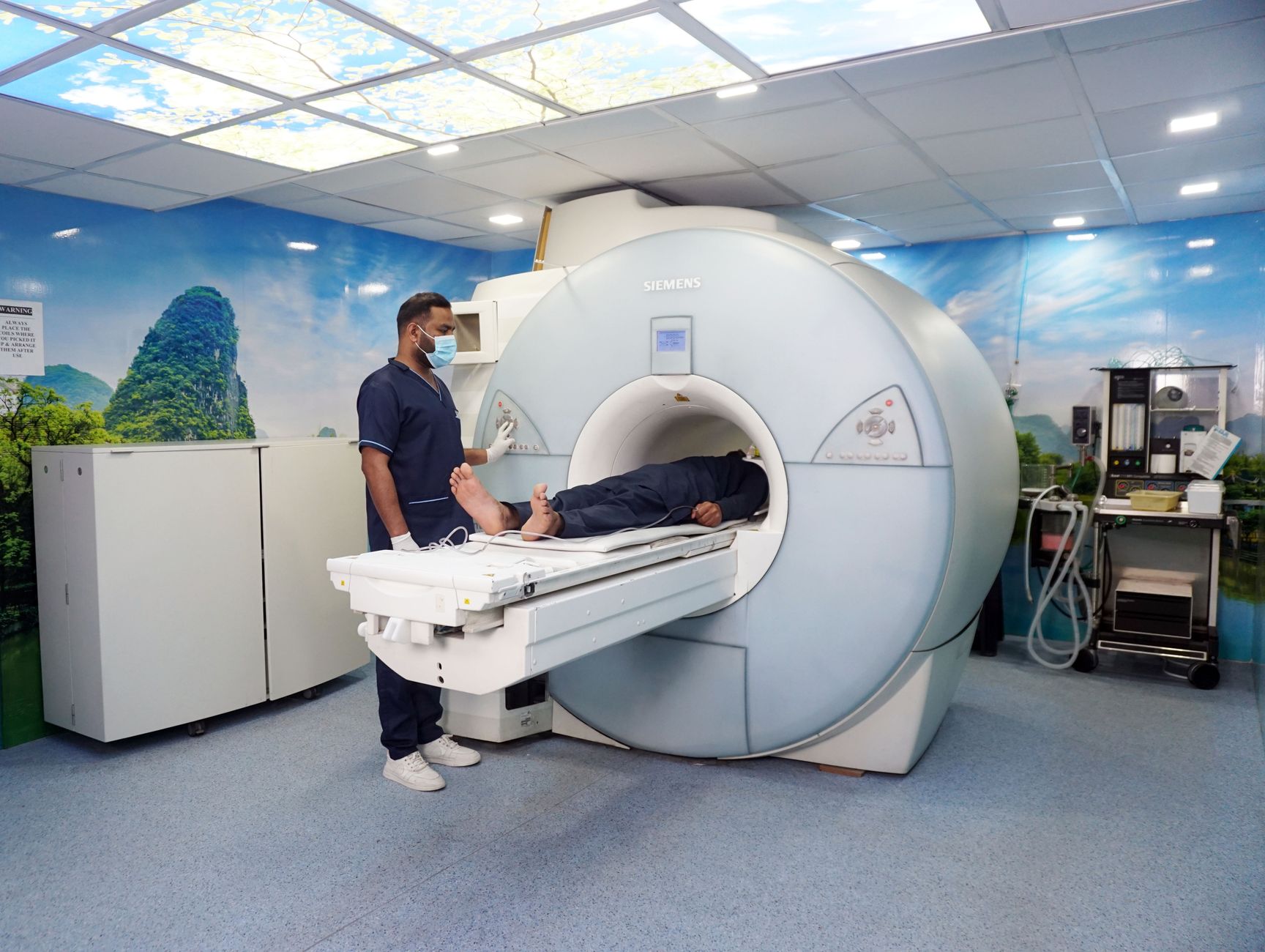
- This advanced AI leverages standard, widely available 3T MRI scans, including historical data, making it immediately applicable across numerous clinical settings without requiring specialized equipment.
- Cortical lesions are strongly linked to cognitive decline and physical disability in MS, and their detection is crucial for understanding disease progression and tailoring effective treatments.
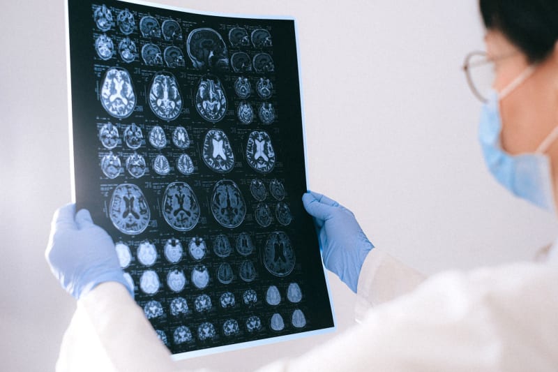
- The AI's ability to detect these subtle lesions, often missed by human radiologists, offers a more comprehensive picture of disease burden and could lead to earlier, more aggressive therapeutic interventions.
- This technology holds the potential to re-evaluate existing MRI archives, uncovering missed diagnoses or providing clearer insights into disease activity for patients already undergoing treatment.
- The development signifies a major leap in neuroimaging for MS, promising to revolutionize how the disease is diagnosed, monitored, and ultimately managed, improving patient outcomes globally.
The Human Face
For individuals living with Multiple Sclerosis, the journey often begins with uncertainty, marked by elusive symptoms and a diagnostic process that can be both lengthy and emotionally taxing. The invisible nature of many MS lesions, particularly those in the cerebral cortex, means that a significant portion of the disease's impact remains hidden from conventional imaging. Patients frequently experience debilitating cognitive and physical symptoms, yet their MRI scans might appear 'normal' or only show a fraction of the true disease burden, leading to delayed diagnoses and suboptimal treatment plans. This new AI offers a beacon of hope, promising to illuminate these hidden aspects of the disease, providing clearer answers and a more accurate understanding of their condition.
Imagine the relief for a patient who has struggled with unexplained cognitive fog or persistent fatigue, only to discover that an AI can now pinpoint the underlying neurological changes that were previously undetectable. This technology doesn't just offer a diagnostic tool; it offers validation and a clearer path forward. It empowers neurologists to initiate more precise and timely interventions, potentially slowing disease progression and preserving quality of life. The human impact extends beyond diagnosis, fostering a sense of understanding and control for those navigating the complexities of MS, moving them from a state of ambiguity to one of informed action.
The psychological toll of living with a chronic, unpredictable disease like MS is immense. The ability to visualize more of the disease's footprint, even retrospectively, can profoundly influence patient management and emotional well-being. It means fewer diagnostic odysseys and more targeted care. Furthermore, for families and caregivers, this enhanced diagnostic clarity can provide much-needed answers and a better understanding of the challenges faced by their loved ones, fostering greater empathy and support. This AI is not just processing data; it's offering a more complete narrative for those whose lives are profoundly shaped by MS.
How We Got Here
The diagnostic landscape for Multiple Sclerosis has long relied on Magnetic Resonance Imaging (MRI) as its cornerstone. However, despite significant advancements in MRI technology over the decades, a critical limitation has persisted: the difficulty in consistently identifying cortical lesions. These lesions, located in the grey matter of the brain, are notoriously subtle and often blend imperceptibly with surrounding healthy tissue on standard MRI sequences, making them challenging even for highly experienced radiologists to detect. This historical blind spot has meant that a crucial aspect of MS pathology, strongly correlated with cognitive impairment and physical disability, has remained largely unquantified in routine clinical practice.
The evolution towards this AI breakthrough began with a deeper understanding of MS pathology and the recognition that cortical lesions play a far more significant role in disease progression than previously appreciated. Researchers have spent years developing specialized MRI sequences, like phase-sensitive inversion recovery (PSIR), which offer improved contrast for cortical grey matter. While these advanced sequences provided better visualization, their widespread adoption was hampered by longer scan times, increased complexity, and the need for specialized equipment or expertise, limiting their utility in standard clinical settings. The challenge was to extract this crucial information from more common, readily available MRI data.
The current breakthrough represents a convergence of advanced neuroimaging research and the explosive power of artificial intelligence. By training sophisticated deep learning models on vast datasets of both standard and specialized MRI scans, researchers at the University of Zurich and ETH Zurich have taught an AI to recognize the subtle, previously imperceptible patterns indicative of cortical lesions on conventional 3T MRI images. This AI effectively 'sees' what human eyes, even with years of training, often miss. This innovative approach bypasses the need for new scanning protocols, directly enhancing the diagnostic yield of existing MRI infrastructure and opening up the possibility of re-evaluating historical patient data with unprecedented accuracy.
Why This Cannot Be Ignored
The inability to reliably detect cortical lesions has been a significant impediment in the accurate diagnosis and comprehensive management of Multiple Sclerosis. These lesions are not merely incidental findings; they are profoundly linked to the most debilitating aspects of the disease, including progressive cognitive decline, memory impairment, and increased physical disability. By missing these crucial indicators, clinicians have been operating with an incomplete picture of a patient's disease burden, potentially leading to delayed or inadequate treatment strategies. This AI's capacity to reveal these hidden lesions fundamentally alters our understanding of individual disease activity and progression, demanding immediate attention from the neurological community.
This technology holds immense potential to revolutionize early intervention strategies. Detecting cortical lesions earlier means that clinicians can initiate disease-modifying therapies (DMTs) more promptly, potentially slowing the accumulation of irreversible neurological damage. For a disease where every moment counts in preserving brain function and preventing disability, this is a game-changer. Furthermore, a more accurate assessment of lesion load, including cortical lesions, can guide personalized treatment decisions, allowing neurologists to select the most effective therapies for each patient, moving beyond a one-size-fits-all approach. Ignoring this advancement would mean perpetuating a less precise and less effective standard of care for MS patients globally.
Beyond individual patient care, the implications for research and drug development are profound. With a more robust method for identifying and quantifying cortical lesions, researchers can better understand the underlying mechanisms of MS, identify new therapeutic targets, and more accurately assess the efficacy of novel treatments in clinical trials. This AI provides a standardized, objective metric that can accelerate the pace of discovery and bring more effective therapies to market faster. The ability to re-analyze vast archives of existing MRI data also presents an unparalleled opportunity to glean new insights into long-term disease trajectories and treatment responses, transforming our collective knowledge of MS.
Possible Paths Forward
The immediate path forward involves the rapid integration of this AI model into clinical practice. Given that it operates on standard 3T MRI scans, its deployment can be relatively swift, requiring software implementation rather than hardware upgrades. Neurological centers and hospitals should prioritize pilot programs to validate the AI's efficacy in diverse patient populations and clinical workflows. This initial phase will be crucial for refining the model, developing robust training protocols for clinicians and radiologists, and establishing best practices for incorporating this enhanced diagnostic capability into routine patient management. The goal is to move from research breakthrough to widespread clinical utility as efficiently as possible, ensuring that patients can benefit from this innovation without undue delay.
Another critical avenue is the retrospective analysis of existing MRI datasets. Hospitals and research institutions possess vast archives of historical MRI scans from MS patients, some spanning decades. Applying this AI to these legacy images could unlock an unprecedented wealth of information, allowing researchers to track disease progression with newfound precision, identify early markers of severe disease, and even re-evaluate the effectiveness of past treatments. This retrospective analysis could provide invaluable insights into the natural history of MS and the long-term impact of various therapeutic interventions, potentially identifying patients who might have been misdiagnosed or undertreated in the past, offering them a chance for re-evaluation and improved care.
Looking ahead, the development of this AI should serve as a catalyst for further innovation in neuroimaging and personalized medicine. Future research should focus on integrating this cortical lesion detection with other biomarkers, such as blood tests or advanced functional MRI, to create even more comprehensive prognostic models. Furthermore, efforts should be made to adapt and refine the AI for lower-field MRI scanners, expanding its accessibility to regions with less advanced medical infrastructure. The ultimate vision is a future where every MS patient receives a precise, early diagnosis and a truly personalized treatment plan, guided by the most complete picture of their disease pathology, fostering a new era of proactive and effective MS management.
Questions People Are Actually Asking
What to Watch
- Monitor for regulatory approvals from health authorities (e.g., FDA, EMA) for clinical use of this AI model, as this will dictate its widespread adoption in diagnostic workflows.
- Observe the rollout of pilot programs and early clinical implementations in major neurological centers, which will provide crucial real-world data on its effectiveness and integration challenges.
- Look for further research validating the AI's performance across diverse patient demographics and different MRI scanner manufacturers, ensuring its generalizability and robustness.
- Track the development of standardized protocols for reporting cortical lesion findings from AI analysis, ensuring consistency and clarity in diagnostic reports for clinicians.
- Pay attention to how this technology influences clinical trial design for new MS therapies, particularly in assessing drug efficacy in reducing cortical lesion burden and cognitive decline.
- Watch for discussions and guidelines from professional neurological societies regarding the integration of AI-powered cortical lesion detection into updated MS diagnostic and monitoring criteria.














Comments
No comments yet. Be the first to comment!